What is the cornea and why does its shape matter?
The cornea provides approximately 70% of the eye's total focusing power. It achieves this because it has a precise, smooth, roughly spherical shape. When this shape is distorted, whether by thinning, bulging, or scarring, the light entering the eye focuses incorrectly and at multiple points, creating the characteristic blurry and distorted vision of ectasia that no flat lens can fully compensate for.
Types of corneal ectasia
The term covers several distinct conditions:
Keratoconus
The most common ectasia, affecting approximately 1 in 2,000 people. Characterised by central or paracentral thinning and a forward bulge, typically beginning in the teenage years. Often bilateral (both eyes), though usually asymmetric.
Pellucid Marginal Degeneration (PMD)
A rarer condition where thinning occurs at the inferior periphery of the cornea rather than centrally. Often misdiagnosed as keratoconus, the distinction matters because lens fitting strategies differ.
Post-refractive surgery ectasia
Can occur after LASIK or other refractive surgeries that remove corneal tissue. The weakened cornea progressively bulges under normal intraocular pressure. Prevention requires careful pre-operative screening; treatment once established is non-surgical.
What does corneal ectasia feel like?
Symptoms progress gradually and include: vision that blurs despite a recent prescription update; monocular double vision or ghosting (a second image seen with one eye closed); increasing sensitivity to glare and halos at night; and the sensation that lenses and glasses 'never quite work'. Because the progression is slow, many patients adapt to worsening vision without realising how much has changed. Topography scans compared over time reveal the changes clearly.
Can ectasia be stopped?
Corneal crosslinking (CXL) is the primary intervention for halting progression. It uses UV light and riboflavin to strengthen the collagen cross-links within the cornea, stiffening it and typically halting or significantly slowing the ectatic process. CXL does not restore lost vision, it stabilises what remains. Once stabilised, specialty contact lenses are the main tool for visual rehabilitation. In severe, late-stage cases where lens fitting is no longer adequate, corneal transplant may be considered.
Non-surgical visual rehabilitation
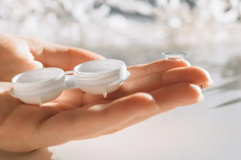
Once ectasia is stable (either naturally or post-CXL), specialty contact lenses are the most effective way to restore functional vision. The lens type depends on the severity and topographic pattern:
RGP and Rose-K lenses
Appropriate for moderate ectasia with a well-defined cone. The rigid lens creates a regular optical surface over the irregular cornea.
Scleral lenses
The gold standard for moderate to severe ectasia, and the first choice for post-surgical ectasia with unusual corneal topography. The complete vaulting over the cornea, combined with the fluid reservoir, provides stable, high-quality vision regardless of the corneal surface.
The role of glasses in ectasia
Glasses remain useful in early ectasia when the irregularity is mild. As the condition progresses, the irregular astigmatism becomes too complex for spectacle correction to adequately address, glasses improve vision but leave it well short of functional. At this stage, specialty lenses become the primary correction tool. It's worth noting that glasses cannot worsen ectasia, but they also cannot slow it, so they are purely a visual aid rather than a management strategy.
Ectasia management at Second Sight, Jaipur
We specialise in the non-surgical visual rehabilitation of corneal ectasia, both spontaneous (keratoconus, PMD) and post-surgical. Optom. Ashima Bhargava has extensive experience fitting Rose-K, RGP, and scleral lenses on ectatic corneas of varying severity. We work in parallel with corneal surgeons when crosslinking is indicated, and coordinate lens fitting once the cornea is stable. If you've been told your vision 'can't be improved' with ectasia, a specialist lens assessment is worth pursuing before accepting that conclusion.
Book a Free Eye Test →